【人気ダウンロード!】 e coli under microscope oil immersion 201104-How to do oil immersion microscopy
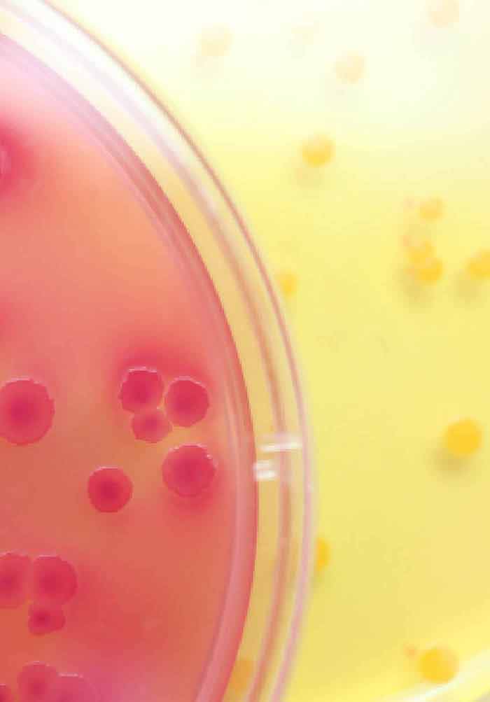
A regular compound microscope will work on 40x, or 100x with an oil immersion lens Just be careful with when you culture the E Coli so no one gets contaminated and use safranin to stain the cells since E Coli is Gram negativeA Betahemolytic colonies of Staphylococcus aureus on sheep blood agar Cultivation 24 hours, aerobic atmosphere, 37°C B Yellow colored colonies of Staphylococcus aureus on Tryptic Soy Agar Carotenoid pigment staphyloxanthin is responsible for the characteristic golden colour of S aureus colonies This pigment acts as a virulence factorPick up a "tiny" amount of an Escherichia coli colony refer to the microscope focusing procedure described in (oilimmersion lens) a Rotate the nosepiece to the empty slot between the 40X and 100X objectives b Add a drop of oil to slide where the light passes through The oil has the same refractive index as
The Oil Immersion Lens Needed To View Bacteria
How to do oil immersion microscopy
How to do oil immersion microscopy-Figure 2 (a) Oil immersion lenses like this one are used to improve resolution (b) Because immersion oil and glass have very similar refractive indices, there is a minimal amount of refraction before the light reaches the lens Without immersion oil, light scatters as it passes through the air above the slide, degrading the resolution of theFirst, by using Escherichia coli (Ecoli), we start at 4x objective, followed by low power 10x objective viewing and the specimen looked clearer at 40x magnificationThe shaped of Ecoli is rodshaped and it is a gramnegative bacteriaThe gram stain of some specimen either gramnegative or grampositive can be resulted by looked at their color whether the bacteria are pink or red for gram


Biol 230 Lab Manual Lab 1
A Betahemolytic colonies of Staphylococcus aureus on sheep blood agar Cultivation 24 hours, aerobic atmosphere, 37°C B Yellow colored colonies of Staphylococcus aureus on Tryptic Soy Agar Carotenoid pigment staphyloxanthin is responsible for the characteristic golden colour of S aureus colonies This pigment acts as a virulence factorPrepared microscope slide of Escherichia coli, bacilli, smear Description This slide of a smear of E coli is best viewed under a microscope with an oilimmersion objective Qty 1 slideA Betahemolytic colonies of Staphylococcus aureus on sheep blood agar Cultivation 24 hours, aerobic atmosphere, 37°C B Yellow colored colonies of Staphylococcus aureus on Tryptic Soy Agar Carotenoid pigment staphyloxanthin is responsible for the characteristic golden colour of S aureus colonies This pigment acts as a virulence factor
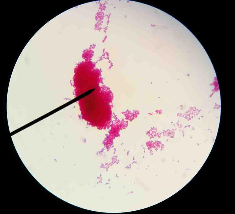
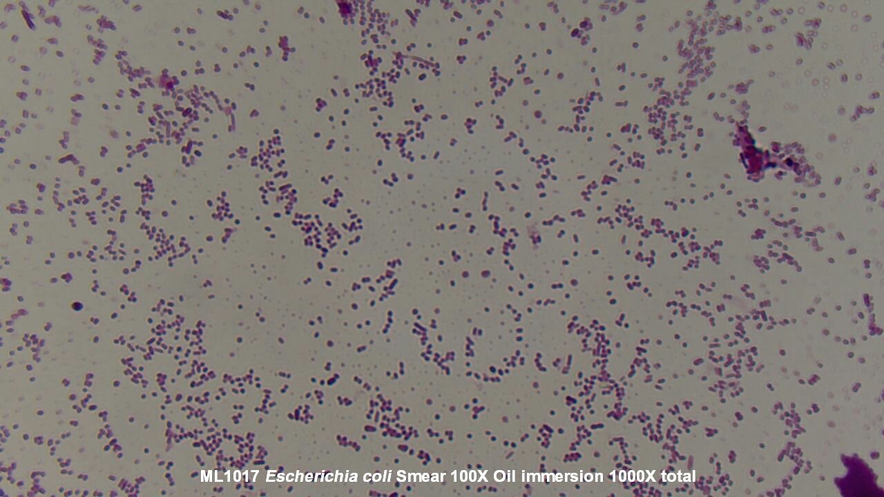
2 Morphology and Staining of Escherichia Coli E coli is Gramnegative straight rod, 13 µ x 0407 µ, arranged singly or in pairs (Fig 281) It is motile by peritrichous flagellae, though some strains are nonmotile Spores are not formed Capsules and fimbriae are found in some strains 3 Cultural Characteristics of Escherichia ColiIn light microscopy, oil immersion is a technique used to increase the resolving power of a microscopeThis is achieved by immersing both the objective lens and the specimen in a transparent oil of high refractive index, thereby increasing the numerical aperture of the objective lens Without oil, light waves reflect off the slide specimen through the glass cover slip, through the air, andLastly, a drop of oil was added to the slide so the sample could be viewed under the microscope using the oil immersion lens Results The E coli had a Gram Stain reaction color of pink and classified as Gramnegative
Browse e coli 100x with oilimmersion pictures, photos, images, GIFs, and videos on PhotobucketThe M Luteus,under oil immersion were purple cocci The mixture bacteria under oil immersion showed a mixture of the two species It was much more difficult to find the E Coli Acid Fast The M Smegmatis under oil immersion appeared reddish, and were rod shaped bacilli The S Aureus under oil immersion appeared blue, and were round cocciBacteria species E coli and S aureus under the microscope with different magnifications Bacteria are among the smallest, simplest and most ancient living


Gram Staining Principle Procedure And Results Learn Microbiology Online


Www Mccc Edu Hilkerd Documents Bio1lab3 Exp 4 000 Pdf
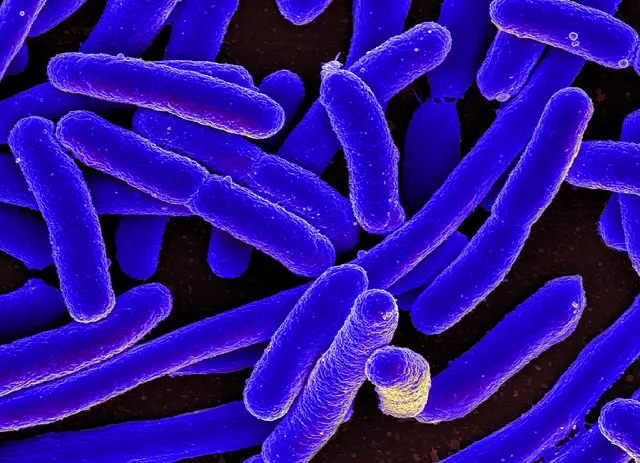
In light microscopy, oil immersion is a technique used to increase the resolving power of a microscopeThis is achieved by immersing both the objective lens and the specimen in a transparent oil of high refractive index, thereby increasing the numerical aperture of the objective lens Without oil, light waves reflect off the slide specimen through the glass cover slip, through the air, andThe prepared microscope slide image of Bacillus Subtilis at left was captured at 400x magnification Learn more here E Coli E Coli under the microscope at 400x E Coli (Escherichia Coli) is a gramnegative, rodshaped bacterium Most E Coli strains are harmless, but some serotypes can cause food poisoning in their hostsEscherichia coli or Ecoli is a gramnegative species of bacillus shaped bacteria that can be easily observed under a microscope, even for those with the untrained eye This bacteria has a fast growth rate, doubling every minutes, making them a common choice for bacterial related research purposes


Mic Uk Observing Bacteria


Laboratory Perspective Of Gram Staining And Its Significance In Investigations Of Infectious Diseases Thairu Y Nasir Ia Usman Y Sub Saharan Afr J Med
Immersion oil onto the part of the slide directly under the lens Now, using the fine adjuster ONLY, focus in onto your organism Those of you using E coli will see something like thisWhen you are through, be sure the microscope is put away properly (ie, all oil wiped off, 10X objective lens in place, stage centered) It is recommended that you keep the slide (To remove immersion oil from smears, place a few pieces of lens paper on the slide to absorb the oil Then, add several drops of xylol to the lens paperE Remember to adjust the light each time the magnification is changed if the area observed is too dark IX 100X Oil Immersion Procedure Immersion oil is used with the 100X objective because it increases the resolution The oil should come in contact with both the lens of the 100X objective and the slide/coverslip


Microscope World Blog Bacillus Subtilis Under The Microscope


Asmscience Examination Of Gram Stains Of Urine
1 Before you plug in the microscope, turn the light intensity control dial on the righthand side of the microscope to 1 Now plug in the microscope and turn it on 2 Place a rounded drop of immersion oil on the area of the slide that is to be observed under the microscope, typically an area that shows some visible stainPlace the slide in the slide holder and center the slide using theColi in a sample Coli microscopy to determine whether a strain s of e Aureus under the microscope with different magnifications Escherichia coli abgekurzt e Coli and related bacteria constitute about 01 of gut flora and fecal oral transmission is the major route through which pathogenic strains of the bacterium cause diseaseTo observe E coli with any detail, you will need to use the 100X lens, which is also known as an oil immersion lens This is the longest, most powerful and most expensive lens on the microscope, requiring extra care when using it As the name implies, the 100X lens is immersed in a drop of oil on the slide


B Cereus 100x With Oil Immersion Oils Immersion Biology


9 Gram Staining Best Practices Microbiologics Blog
During the heating step of the acidfast stain, the carbol fuchsin is allowed to come to a boil and dry out The bacteria are red after completion of the staining process when viewed under oil immersion, which means the bacteria are acidfast positive (end of exp 10)Inclusion bodies in E coli Tuner (D) pLacI, as observed under 100X magnification of oil immersion microscope 313 104 The Coomassie blue stained polyacrylamide gel analysisLastly, it is examined under the oil immersion objective and picture was taken Results A) Direct Staining with Basic Dyes Escherichia coli Figure 1 Escherichia coli stained with crystal violet under the microscope with magnification of 100X


The Oil Immersion Lens Needed To View Bacteria


Gram Stain
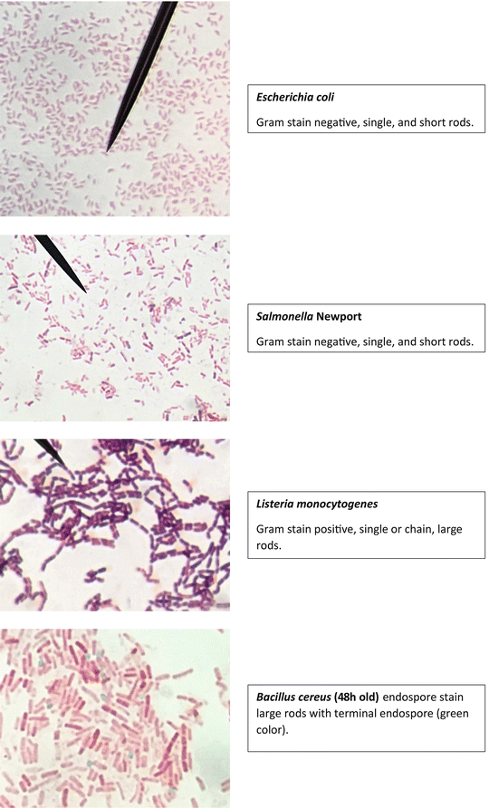
An alternate focusing technique is to first focus on the slide with the yellowstriped 10X objective by using only the coarse focus control and then without moving the stage, add immersion oil, rotate the whitestriped 100X oil immersion objective into place, and adjust the fine focus and the light as needed This procedure is discussed in the Introduction to the lab manualFinally, add a drop of immersion oil directly to the slide, and then, examine the slide using a light microscope with a 100X oil objective To perform endospore staining, first, prepare a 05% malachite green solution by mixing 0 125 grams of malachite green powder with 25 milliliters of distilled water, and then vortex the solution until(5) A prepared slide of Bacillus cereus was observed with the microscope under 1000X magnification using oil immersion This organism had green dots located in the center of the cell which indicated that had nutrient depletion in the Bacillus cereus had caused it to produce coccobacillus shaped spores in order to survive


Biol 230 Lab Manual Lab 1


Photomicrographs
Lastly, it is examined under the oil immersion objective and picture was taken Results A) Direct Staining with Basic Dyes Escherichia coli Figure 1 Escherichia coli stained with crystal violet under the microscope with magnification of 100XSince E coli are about 2 micrometres long, if you can't discriminate between two points that a four microns apart with your lens, you going to see a blurry smudge if you see anything at all It is routinely possible, using highend, oil immersion 100x lenses microscopes, to get down to 02 micrometre resolution, which is smaller than an E coliGram stained smear, 100X (oil immersion) We and our partners process personal data such as IP Address, Unique ID, browsing data for Use precise geolocation data Actively scan device characteristics for identification Some partners do not ask for your consent to process your data, instead, they rely on their legitimate business interest


Aph162 Report 1


Biol 230 Lab Manual Lab 1
Put a drop of immersion oil directly on each of the three bacterial smears on your slide, then switch to the oil immersion lens Image Microscope objective lenses, T Port From the Virtual Microbiology Classroom on ScienceProfOnlinecom Observing bacteria under oil immersion Don't EVER use coarse focus when working with high dry or oilNotice that the cells appear much smaller than in the image that has Ecoli with green color added This image was generated using a light microscope which typically magnifies Jennifer Julizar BIOL 351 Section 906 Staining Results and Conclusions using microscope under 1000X magnification with oil immersionColi in a sample Coli microscopy to determine whether a strain s of e Aureus under the microscope with different magnifications Escherichia coli abgekurzt e Coli and related bacteria constitute about 01 of gut flora and fecal oral transmission is the major route through which pathogenic strains of the bacterium cause disease


Photomicrographs


E Coli Gram Stain Page 1 Line 17qq Com
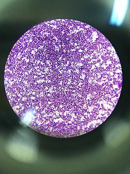
Lastly, a drop of oil was added to the slide so the sample could be viewed under the microscope using the oil immersion lens Results The E coli had a Gram Stain reaction color of pink and classified as Gramnegative6 Blot dry with KimWipe and observe under microscope using oil immersion lens (100x) E coli is a Gram negative rod S aureus is a Gram positive coccus Exercise 9 The AcidFast Stain 1 Observe demonstration slide or #25 on Microbiological Chart of Mycobacterium tuberculosis 2 Specify colors of nonacidfast versus an acidfast organismLarge, beige, drylike colonies Escherichia coli Small, pinpoint or dotlike, white colonies Bacteria E coli Must observe under 400x Very small & motile Must be viewed under oilimmersion Allows you to see Shape Size


2 3 Instruments Of Microscopy Microbiology Canadian Edition


Toxins Free Full Text Identification Of Three Type Ii Toxin Antitoxin Systems In Streptococcus Suis Serotype 2 Html
E Coli Under The Microscope Types Techniques Gram Stain Gram Stained Bacterial Strain 68 Observed Under The Nikon Eclipse How To View Bacteria Through Microscope With Oil Immersion Hd00 21a Colony Of Bacteria Under A Microscope Magnification OfBacteria Under the Microscope Types, View under the microscope starting with low power (for high power, add immersion oil) Observation (Discussion) Depending on the sample under investigation, students will have the opportunity to observe and identify the size and shape of the bacteria Like E coli live in the intestines of animalsFocus the 100xTM low power objective lens on the smear and examine Cover the 400xTM objective lens with a finger cot to prevent this lens from coming in contact with the oil Place a drop of immersion oil directly on the sample Turn nosepiece until 1000xTM objective lens snaps into position


E Coli Gram Stain Page 1 Line 17qq Com


Microbiology Lab Exercise 6 Acid Fast Staining Flashcards Cram Com
Large, beige, drylike colonies Escherichia coli Small, pinpoint or dotlike, white colonies Bacteria E coli Must observe under 400x Very small & motile Must be viewed under oilimmersion Allows you to see Shape SizeBacteria Under the Microscope Types, View under the microscope starting with low power (for high power, add immersion oil) Observation (Discussion) Depending on the sample under investigation, students will have the opportunity to observe and identify the size and shape of the bacteria Like E coli live in the intestines of animalsTo observe E coli with any detail, you will need to use the 100X lens, which is also known as an oil immersion lens This is the longest, most powerful and most expensive lens on the microscope, requiring extra care when using it As the name implies, the 100X lens is immersed in a drop of oil on the slide


Acid Fast Stain Free Microbiology Images From Science Prof Online


Laboratory Perspective Of Gram Staining And Its Significance In Investigations Of Infectious Diseases Thairu Y Nasir Ia Usman Y Sub Saharan Afr J Med
The glass slides were washed in PBS 3 times, and the dyed bacteria were observed under the oil immersion lens of the microscope Growth Characteristics E coli Kac LT(S63K) Δ STb and C902 were inoculated into liquid LB medium (pH = 72) and cultured overnight at 37°C1 Before you plug in the microscope, turn the light intensity control dial on the righthand side of the microscope to 1 Now plug in the microscope and turn it on 2 Place a rounded drop of immersion oil on the area of the slide that is to be observed under the microscope, typically an area that shows some visible stainPlace the slide in the slide holder and center the slide using theAdd oil immersion to the stained area and observe it under the microscope using a 100X objective India ink method is a type of negative staining method, which stains both the bacterial cell and its background (but not a capsule) As a result, a capsule appears as a bright halo between the violet bacterial cell and a darker background


What Does An E Coli Bacteria Look Like Under A Microscope Quora


Lab 1
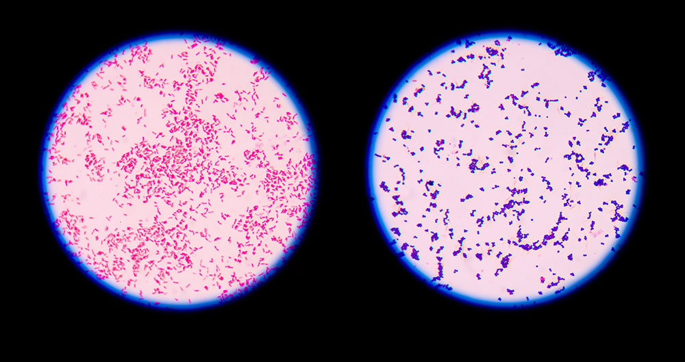
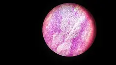
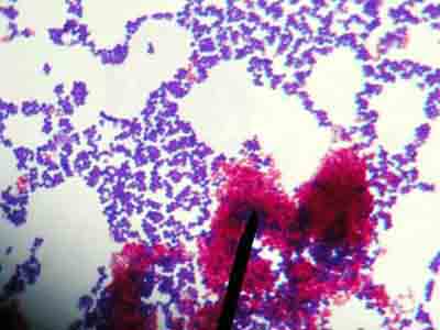
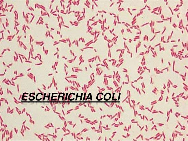
When viewed under the microscope, Gramnegative E Coli will appear pink in color The absence of this (of purple color) is indicative of Grampositive bacteria and the absence of Gramnegative E Coli Escherichia coli under 10х90х magnification using fuchsine as a dye by ElNokko (Own work) CC BYSA 40 (http//creativecommonsorg/licenses/bysa/40), via Wikimedia CommonsBacteria Under The Microscope E Coli And S Aureus Youtube Academic Oup Com Cid Article Pdf 65 4 544 Cix356 Pdf Pathogenic E Coli Microscopic Field Oil Immersion Objective Showing The Accumula Cocci Bacteria In Urine Urinary Tract Infection Pathology BritannicaLastly, a drop of oil was added to the slide so the sample could be viewed under the microscope using the oil immersion lens Results The E coli had a Gram Stain reaction color of pink and classified as Gramnegative


Escherichia Coli Bacteria E Coli Stock Footage Video 100 Royalty Free Shutterstock


Why Would You Need Microscope Immersion Oil And How To Use It
6 Blot dry with KimWipe and observe under microscope using oil immersion lens (100x) E coli is a Gram negative rod S aureus is a Gram positive coccus Exercise 9 The AcidFast Stain 1 Observe demonstration slide or #25 on Microbiological Chart of Mycobacterium tuberculosis 2 Specify colors of nonacidfast versus an acidfast organism


Lab 1


Www Lycoming Edu Schemata Pdfs Fritz Bacterial growth Fall14 Pdf


Biol 230 Lab Manual Lab 1


Staphylococcus Aureus Under Microscope Microscopy Of Gram Positive Cocci Morphology And Microscopic Appearance Of Staphylococcus Aureus S Aureus Gram Stain And Colony Morphology On Agar Clinical Significance


E Coli Under The Microscope Types Techniques Gram Stain Hanging Drop Method


Microbiology Lab Exercise 10 Smear Preparation Exercise 11 Simple Staining Ex 12 Negative Staining Review Lab Results From Last Week Ex 8 Aseptic Technique Did You Successfully Get Bacteria Growing In Your Broth And Slant Ex 6 The


Gram Positive And Gram Negative Bacteria Structures Characterisitics


E Coli Under The Microscope Types Techniques Gram Stain Hanging Drop Method


What Does An E Coli Bacteria Look Like Under A Microscope Quora


Lab 1


Photomicrographs


Microscopy For The Winery Viticulture And Enology


Meningitis Lab Manual Primary Culture And Presumptive Id Cdc


Staphylococcus Aureus Under Microscope Microscopy Of Gram Positive Cocci Morphology And Microscopic Appearance Of Staphylococcus Aureus S Aureus Gram Stain And Colony Morphology On Agar Clinical Significance


Bacteria Under The Microscope E Coli And S Aureus Youtube


Slide Escherichia Coli


Staining Technology And Bright Field Microscope Use Springerlink


Microscopy And Staining


Photomicrographs


Differential Staining Techniques Microbiology A Laboratory Experience


Endospore Staining Principle Procedure And Results Learn Microbiology Online
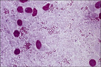


Bacterial Pathogens Microscopy Culture And Identification Veterian Key


Microscope Slide An Overview Sciencedirect Topics


Q Tbn And9gcs3nlev0tx1pfsddwm6y9ajujk4lxjzmg7ksdxctgnipv0l50c Usqp Cau


Http Coltonanderson1 Weebly Com Uploads 2 4 3 0 Manual Pdf


Biosensor Platforms For Rapid Detection Of E Coli Bacteria Intechopen


Acid Fast Stain Free Microbiology Images From Science Prof Online


Morphology Of E Coli Cells Under Microscope At 100 Magnification Download Scientific Diagram


Observing Bacteria Under The Light Microscope Microbehunter Microscopy


Why Would You Need Microscope Immersion Oil And How To Use It


What Does An E Coli Bacteria Look Like Under A Microscope Quora


Acid Fast Stain Free Microbiology Images From Science Prof Online


What Does An E Coli Bacteria Look Like Under A Microscope Quora


Gram Stain


Www Mccc Edu Hilkerd Documents Bio1lab3 Exp 4 000 Pdf


Zkfaa Bioproses Lab 1 Principles And Use Of Microscope


Microscopy Aquarium Advice Aquarium Forum Community


Google Answers Microscopes


Microscopy And Staining


Observing Bacteria Under The Light Microscope Microbehunter Microscopy


Ex 6 Negative Staining Scientist Cindy


Microscopic Field Oil Immersion Objective Showing The Accumula Tion Download Scientific Diagram


Bacillus


What Does An E Coli Bacteria Look Like Under A Microscope Quora


Simple Bacterial Stain Free Microbiology Images Photographs


Bacterial Staining Microbiology Images Photographs And Videos Of Gram Acid Fast Endospore


High Precision Characterization Of Individual E Coli Cell Morphology By Scanning Flow Cytometry Konokhova 13 Cytometry Part A Wiley Online Library


Staphylococcus Epidermidis Under Microscope Microscopy Of Gram Positive Cocci Morphology And Microscopic Appearance Of Staphylococcus Epidermidis S Epidermidis Gram Stain And Colony Morphology On Agar Clinical Significance


Microbe Classification Using Deep Learning By Steven Towards Data Science


Escherichia Coli Bacteria E Coli Stock Footage Video 100 Royalty Free Shutterstock


Biol 230 Lab Manual Lab 1


9 Microbiology Ideas Microbiology Medical Laboratory Medical Laboratory Science


Bacterial Staining Microbiology Images Photographs And Videos Of Gram Acid Fast Endospore


Biol 230 Lab Manual Lab 1


Q Tbn And9gcqkye60ou Johpr02n Mbv1fferrjpdh Lnct7ymdf5qhyia1ld Usqp Cau
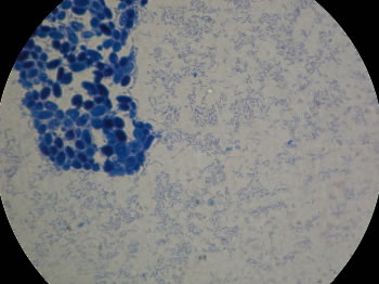


The Virtual Edge


Mic Uk Observing Bacteria
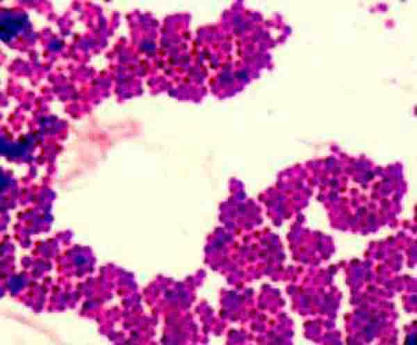


How To View Bacteria Through Microscope With Oil Immersion


Morphology Of Parasites Identified By Light Microscopy In Stool Samples Download Scientific Diagram


Cell Division Of E Coli With Continuous Media Flow Youtube


Pin On E Coli
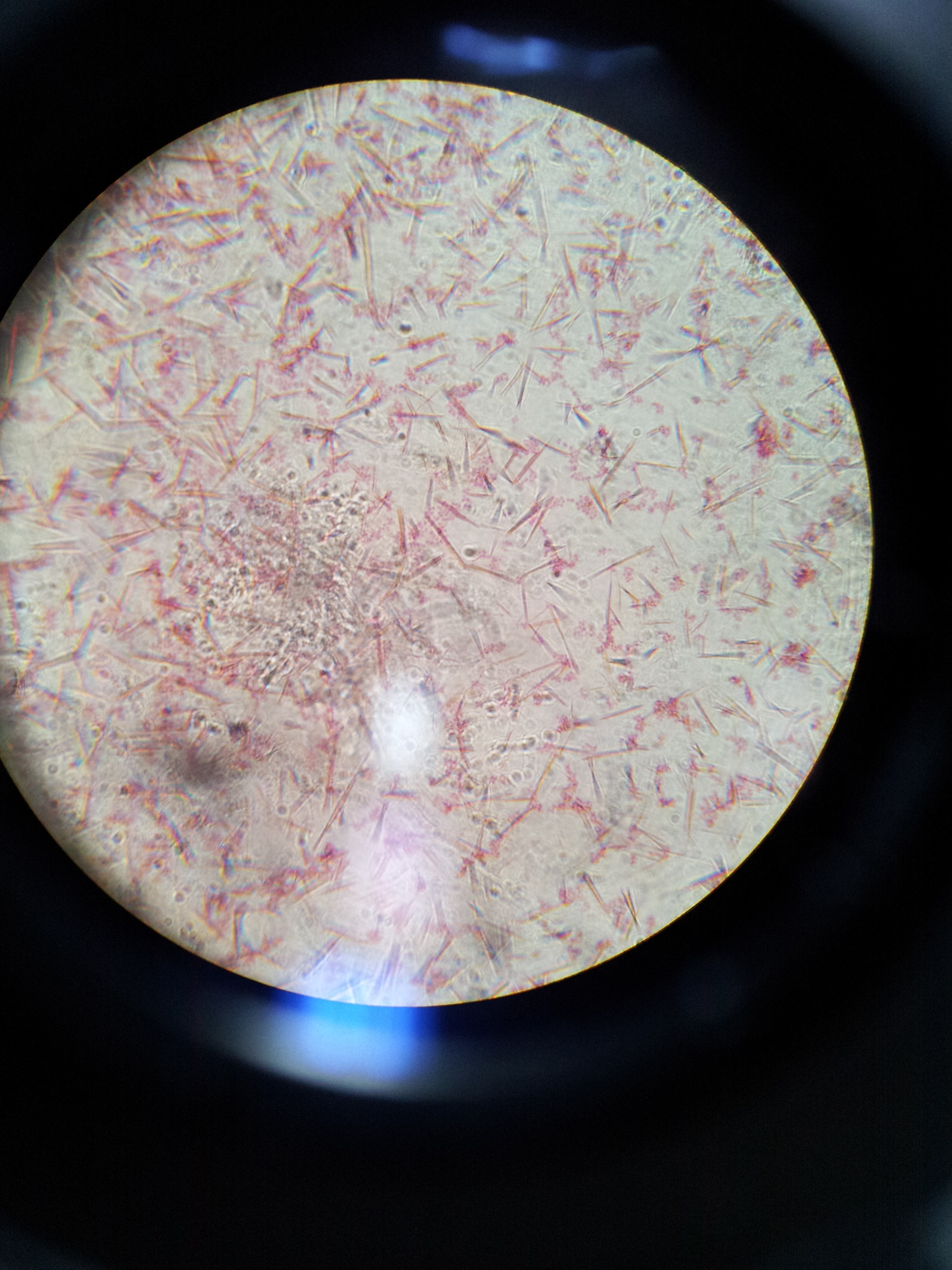


Lab 1 Principles And Use Of Microscope Ibg 102 Lab Reports


Observing Bacteria Under The Light Microscope Microbehunter Microscopy


Biol 230 Lab Manual Lab 1


Endospore Under Microscope 100x Oil Immersion Lens Youtube


What Does An E Coli Bacteria Look Like Under A Microscope Quora


Biol 230 Lab Manual Lab 1
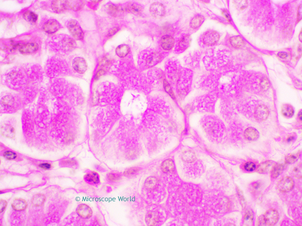


Why Would You Need Microscope Immersion Oil And How To Use It


Asmscience Diagnostic Methods For The Enteroaggregative Escherichia Coli Infection


Q Tbn And9gctqlwezc G Rsexb5gmw Uv65za98k6p92dhlvblkp4mnrofyo Usqp Cau


Differential Staining Techniques Microbiology A Laboratory Experience


E Coli Gram Stain Page 1 Line 17qq Com


Gram Stain


Lab 2 Lab 3


Biol 230 Lab Manual Lab 1


Gram Stain


E Coli Gram Stain Introduction Principle Procedure And Result Interpret


Q Tbn And9gcq Fnvqgh9s1fp Ssci5dy6dtinzl2mp33u1pncpzysdndo9cmk Usqp Cau


コメント
コメントを投稿